
 |  |
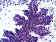 | 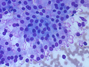 |
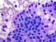
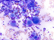
| The thyroid web atlas contains close to 300 images of key thyroid lesions in an easy-to-search format. All images have been independently reviewed and approved by the committee members. Although the majority of the images presented here are from direct smears of an FNA sample, there are numerous examples of liquid-based preparations as well, illustrating the key morphologic differences. Emphasis has been placed on presenting “classic’ examples of common thyroid lesions, but images from uncommon and rare diseases are included as well to make this as comprehensive as possible. The images are formatted to conform to the print atlas “The Bethesda System for Reporting Thyroid Cytopathology” (Ali SZ, Cibas ES, eds, Springer, 2010). The images are tagged with the keyword (entity name) and are also searchable by chapter index and preparation type (direct-smear versus liquid-based). |
Using the Image Atlas
- Click on an album to see the thumbnail images
- Click on the thumbnails to see the intermediate images
- Click on the intermediate image to obtain the full sized image
These images are intended for educational purposes and may be freely used for such as long as the Papanicolaou Society and the Bethesda System for Reporting Thyroid Cytopathology are credited.