Case of the Month ...
Case History
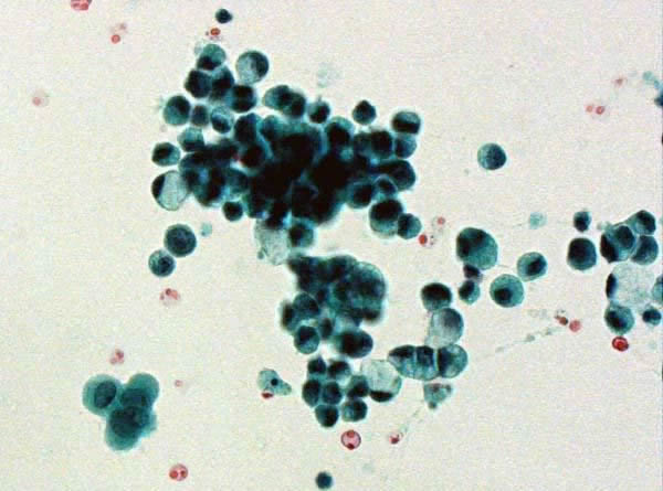
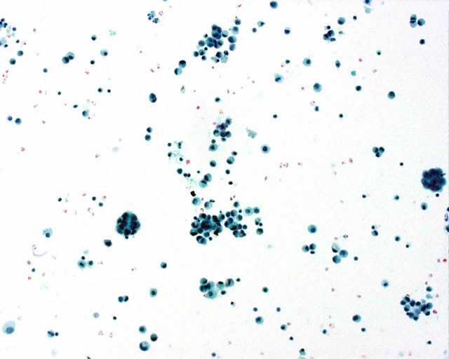
A 48-year-old woman presented with early satiety and increased abdominal girth. Ascitic fluid was removed and sent for cytologic evaluation. The first two Papanicolaou stained cytospins demonstrate a cellular fluid with medium sized discohesive cells. Some cytoplasmic vacuoles are present and most of the bland nuclei are eccentrically located. The Diff Quik stain shows cells with a mildly increased nuclear to cytoplasmic ratio, smooth nuclear contours and prominent nucleoli.
Diagnosis & Discussion
click on image for larger version
Diagnosis: adenocarcinoma, signet ring cell type.
Discussion: Features that generally aid in classifying a fluid as malignant include a distinct second cell population, cells with a high nuclear to cytoplasmic ration with irregular nuclear contours and prominent nucleoli, cohesive cells in three dimensional clusters with shared borders (as opposed to the hobnailing of mesothelial cell clusters). Signet ring cell adenocarcinoma can be extremely difficult to diagnose in fluids due to the tumor cell homogeneity, similar or smaller size as mesothelial cells, and single cell pattern. Tumors that have a propensity to present as single cells (often referred to as a mesothelial cell pattern) include breast, stomach, lymphoma, melanoma and signet ring cell adenocarcinomas such as from the stomach, breast, and pancreas. In this case, the malignant cells are so numerous that the distinct second cell population is difficult to discern. In the second Papanicolaou stained slide, mesothelial cells can be appreciated in the lower left-hand corner to contrast with the remaining tumor cells. The cells of signet ring carcinoma resemble histiocytes with the irregular nuclear contours and vacuolated cytoplasm. However, the number of cells without other prominent inflammatory cells would be unusual for histiocytes. In addition, most of the nuclei are eccentric due to compression by the cytoplasmic mucin. This latter feature can be helpful as a PAS with diastase stain should highlight the cytoplasmic mucin. Mesothelial cells and histiocytes occasionally have focal cytoplasmic mucin but they do not usually form large cytoplasmic globules.
Working through the single cell pattern differential diagnoses, lymphoma is less likely due to the abundant cytoplasm. One of melanoma’s many patterns include plasmacytoid; however, the small size of the cells and lack of intracytoplasmic pigment, intranuclear psuedoinclusions or double nuclei argue against melanoma. There is nothing cytologically that should point the cytologist to a particular site of origin for the signet ring carcinoma. If enough fluid is retained, differential cytokeratins, lung and breast immunochemistry markers may be helpful.
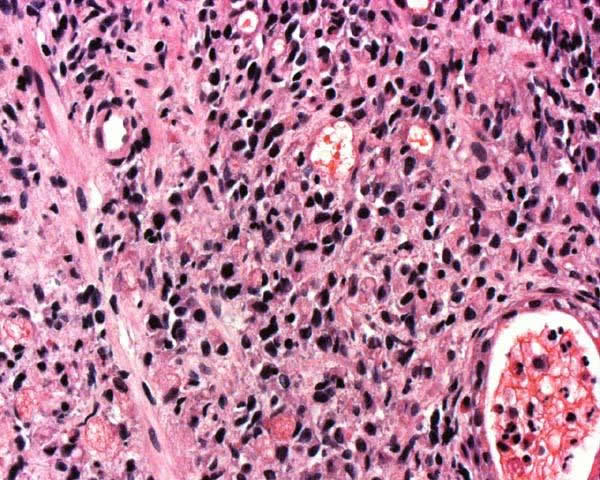
Surgical biopsy correlation: Due to the patient’s early satiety, the gastrointestinal tract was evaluated first for a primary site. Stomach biopsies revealed an adenocarcinoma, signet ring cell type (H and E stain 20x and 40x).